

Tuberculosis (TB) in cattle was first recorded in South Africa late in the eighteenth and early in the nineteenth century. It was most likely brought in by imported animals infected with the disease. Bovine TB is a chronic disease caused by the bacterium Mycobacterium bovis and affects practically all vertebrate animals. It is widespread in developing countries in Africa, parts of Asia and the Middle East.
Economic losses occur due to mortality, poor performance of chronically sick animals, and serious restrictions in international trade. Australia, Denmark, Sweden, Norway, Austria, Switzerland, Canada and several other countries are currently bovine TB-free.
Three types of TB are of economic and health significance:
- Bovine TB, caused by Mycobacterium bovis;
- Human TB, caused by Mycobacterium tuberculosis;
- Avian TB, caused by Mycobacterium avium.
Although related, causal organisms differ and can only be positively differentiated from one another by laboratory tests. Cattle are the primary hosts of M. bovis, although other domesticated and wild mammal species may also become infected. The organism can cause disease in species as diverse as buffalo, kudu, sheep, goats, horses, pigs, cats, dogs, humans and birds.
It is a zoonosis (a disease transmittable from animals to humans). It can also be transmitted from human to animal, although this is rare. Bovine TB was declared a controlled disease in South Africa in 1911 and is classified by the World Organisation for Animal Health as a notifiable animal disease with specific impact on international trade.
Contracting the disease
M. bovis can enter the body through breathing in air via the mouth, or through damaged/cracked skin. The incubation period between infection and symptoms developing usually lasts months. Bovine TB may be present but dormant in an animal for years, only to be activated during periods of high stress or old age. In most cases, transmission from an infected animal with open wounds to a healthy animal is a direct process.
Direct infection
Direct infection of the respiratory system usually occurs in stables or other confined areas. Infected cattle cough out fragments or droplets of infected mucous, transferring the bacteria to other animals or humans breathing in the air in the immediate vicinity. Bacteria may also end up on the ground, wafted upwards in the dust and breathed in.
Oral infection
Oral infection may take place by drinking infected milk or water. Pastures or feed bins contaminated with bacteria originating from open respiratory systems, in which mucous is coughed up and again swallowed, play an important role in infection. The disease can be transmitted by saliva or food particles in the digestive system contaminated by lesions in the digestive tract and voided in the dung, as well as by milk, urine or sheath secretions, should the specific organ be affected. Affected lymph nodes that void externally may also infect food or water sources.
It can also be transmitted iatrogenically via contaminated needles, milking machinery, speculums or other livestock apparatus, or during mating, should the genitals be affected. Infection through wounds cannot be excluded. Transmission to an unborn calf when the uterus is infected or the cow develops general lesions is also possible.
Bacterial survival outside the body
The length of time that the bacteria can remain infective outside the body depends on climatic conditions. Desiccation and direct sunlight is detrimental. It appears that the bacteria can survive a week on veld and several months in a cool (12°C to 24°C), dark and moist environment, if not exposed to direct sunlight. Standing drinking water can remain infective for 18 days; in moist dung, the bacteria can survive for six weeks to eight weeks. Tilling the soil covered with infective dung will reduce the infective period to seven days in warm sunlight.
Infection
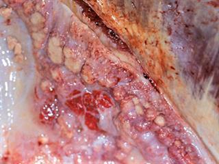
Infection takes place by bacteria entering the body, usually via the lungs. Bacteria may also enter via the digestive system as well as through skin wounds. After establishing themselves, the bacteria stimulate the formation of tumour-like lesions called tubercles. A tubercle grows in size and its centre may die away, become cheese-like and eventually calcify. The colour is light-orange.
Where bacteria are released at the original focus, they may spread to other parts of the body via the lymphatic system or bloodstream to form new tubercles. Should many bacteria end up in the bloodstream, they will spread throughout the body and form lesions, causing toxaemia (blood poisoning). This in turn leads to weakness, lack of energy and eventual death.
Occasionally, lesions may be sufficiently encapsulated by connective tissue to inhibit further spread through the body, limiting the disease.
Lesions also usually develop in the lymph nodes, draining affected body parts or organs. For this reason, the nodes are usually examined to determine infection.
Symptoms
Bovine TB is generally a chronic and at times invisible disease, with symptoms developing months or years after infection.
In most cases, the animal’s lymph nodes are affected, followed by its lungs, udder and other internal organs. Most infected cattle testing positive in the tuberculin test (skin test) exhibit no externally observable disease symptoms.
Animals with scattered lesions in the body may eventually become emaciated, with a variable appetite, fluctuating body temperature and dull hair coat. Such animals become listless, although the eyes remain clear. Signs become more apparent during times of stress and when the body becomes subject to increased demands, such as when calving.
In cases of advanced lung infection, a single suppressed, moist cough may be heard, especially early in the morning, after exercise or when it is cold. Breathing becomes laboured, and there is widespread lung damage. The animal becomes emaciated and develops serious breathing problems. Enlarged lymph nodes in the lungs constrict the air passages, exacerbating the problem. Repeated bloating may occur due to the enlarged lung lymph nodes exerting pressure on the oesophagus.
Excessively enlarged lymph nodes in the digestive system obstruct the digestive tract, leading to diarrhoea and constipation. Enlarged superficial lymph nodes can be seen or felt. Uterus infection (metritis)can either obstruct conception or lead to abortion at advanced pregnancy. Infection via the udder poses a danger for a calf or person drinking infected milk that has not been pasteurised or boiled. It may be difficult to distinguish this type of mastitis from that caused by other organisms.
Bovine TB may often lead to a hardening or enlargement of the upper section of the udder quarters. In such cases, the lymph nodes enlarge. Enlarged lymph nodes without palpable lesions in the udder are also indicative of the disease. The milk initially appears normal, while at a later stage flakes may be seen in milk that has been standing for some time.In an advanced stage, the milk is a light yellow-brown, and watery.
Diagnosis
A vet is responsible for diagnosing and confirming bovine TB in cattle and other animals. History and clinical signs must be taken into consideration.
Diagnostic methods may include the following:
- Smears can be made of fragments of coughed-up mucous, from the solid fraction of centrifuged milk and urine, from sheath secretions, from dung or from affected lymph nodes and other organs. The smears are stained according to the Ziehl-Neelsen method and examined under an optical microscope.
- A biological test can be conducted by injecting a suspension of the abovementioned samples into guinea pigs and conducting a postmortem examination after six weeks.
- The same material can be used to culture bovine TB bacteria in a suitable medium. However, the disadvantage is that, as in the case of the biological test, it takes time to yield results.
- The intradermal bovine tuberculin test is commonly used to trace bovine TB infection in cattle. It is based on a sensitivity that develops in the body after infection with M. bovis. A delayed hypersensitivity reaction, in which a swelling develops in the skin, gives a positive answer. However, several factors may lead to a false negative result.
- The bovine tuberculin test is often combined with the avian tuberculin test for more accurate and reliable results. The living bacteria may cause pathological changes, visible lesions and disease. In South Africa, conducting a tuberculin test is subject to government approval and specific conditions.
- Other tests available include the temperature test, the double intradermal test and the Stormont test. Advanced tests include the polymerase chain reaction (PCR) test or the enzyme-linked immunosorbent assay (ELISA) test.
Bovine TB as a Zoonosis
While rare, humans infected with M. tuberculosis can infect cattle. Although progressive lesions usually do not develop, cattle are nevertheless sensitised and this complicates the interpretation of the tuberculin test. Cases of people with open lesions becoming a source of infection for cattle are on record, and the danger of people infected with M. bovis reinfecting TB-free cattle herds should always be kept in mind.
People usually become infected with M. bovis by drinking unpasteurised infected milk or eating dairy products made from such milk. People can also become infected directly through damaged skin or breathing in infected air, especially in the presence of herds with a high incidence of the disease and in indoor situations such as stables and sheds where there is close contact between cattle and humans.
It should be noted that milk pasteurised at 72°C for 15 seconds, or submitted to the ultra-high temperature (UHT) process, is safe for human consumption. Pasteurising milk kills all pathogenic bacteria, including those causing bovine TB. A few cases of humans becoming infected via wounds such as an accidental cut with a knife during a postmortem examination are on record.
Humans usually develop lesions in the skeleton, mesenteric glands and occasionally lymph nodes. Cases of lung infection have been recorded. Chickens, pigs, sheep, goats and horses, as well as certain wildlife species, including lion and kudu, are also susceptible to bovine TB. Such infected animals pose a health danger to humans. A vet must conduct a confirming postmortem examination on any animal suspected of having been infected.
Economic risk
The production of meat and milk in infected animals is reduced by between 10% and 25%. In a small percentage of cases, the genital organs of both genders may be affected, leading to reproduction losses due to infertility and abortions. A general spreading of the disease throughout the body can lead to mortality. This is more common under conditions of poor nutrition and a shortage of fodder, such as during drought.
Under extensive production conditions, bovine TB usually spreads slowly. However, the modern trend is to farm more intensively, with more animals in a smaller area. This creates favourable conditions for the rapid spread of the disease in stalls, at feeding troughs, at lick bins and watering points, and through mass grazing at high density. Developed countries are placing increasingly more stringent demands on the eradication of bovine TB in the countries from which they import.
Control and prevention
It is in the interest of public health to eradicate bovine TB. In South Africa, bovine TB is a controlled animal disease in terms of the Animal Diseases Act of 1984 and its accompanying regulations. Apart from requiring compulsory notification of the disease, the regulations also make testing compulsory where the disease occurs or is suspected. The regulations also have the power to impose a quarantine or destruction programme.
Several testing schemes, including accreditation and the diagnostic herd testing scheme, have been established to trace, control, combat and eradicate it. Farmers should consult the local state vet about preventing and controlling the disease. No effective vaccine has yet been developed to prevent bovine TB, and treating cattle with anti-microbic compounds is neither practical nor cost-effective.
Email Dr Jan du Preez, managing director of the Institute for Dairy Technology, at [email protected], or Dr Faffa Malan, manager of the Ruminant Veterinary Association of SA, at [email protected].


