

Livestock disease causes South African farmers major economic losses every year. Infectious diseases, tick-borne and insect-borne diseases play a prominent role in these losses. Several diseases are economically significant in dairy cattle worldwide. Mastitis is known to be the most costly disease in dairy farming while enzootic bovine leukosis (EBL) is the most economically important retroviral disease.
Other retroviruses cause jaagsiekte (ovine pulmonary adenocarcinoma) in sheep, equine infectious anemia (EIA) in horses and various diseases in goats. EBL is caused by a retrovirus known as bovine leukaemia virus (BLV). Three different forms are recognised: cattle with no clinical symptoms with antibodies, cattle with persistent white blood cells (leucocytes) and cattle that develop solid lymphoid tumours.

Dr Jan du Preez
The small percentage of animals that develop fatal cancer can incubate the virus for a long period. EBL is not associated with sporadic bovine leukosis (SBL) that occurs primarily in young cattle from 4 months to 2 years old and mainly affects the skin and thymus gland. Both EBL and SBL occur in Southern Africa.
Distribution and occurrence EBL occurs worldwide, although infections are more prevalent in Eastern Europe, the Americas, Australia and Africa. Annual economic losses due to having to destroy infected animals, reduced milk production and reduced exports amount to several million rand.
The BLV infection rate in herds varies from 1% to 100% and annual mortality in severely infected dairy herds is estimated at 5%. All infected cattle develop antibodies and test positive for BLV. Only 30% develop a persistent lymphocytosis (a certain type of white blood cell) and of these only 30% develop tumours. Once infected, cattle remain infected for life.
EBL widespread
Holstein-Friesian, Jersey, other dairy cattle breeds and cross-bred dairy cattle have antibodies against BLV, indicating that infection occurred in the past. More than 50% of the animals in some dairy herds are infected, but the infection rate in beef herds is very low. EBL in South Africa is widespread especially in the Eastern Cape, Western Cape and Mpumalanga. There is no official information on the extent and impact of EBL in South Africa, and very little information from other African countries.
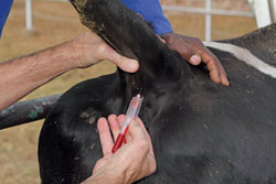
Use a separate sterilised needle for each animal when drawing blood samples for diagnostic tests or injecting.
How does infection occur?
The DNA of the virus is closely associated with cells in blood and milk. It is directly transferred between blood cells of infected and healthy animals. This transfer occurs commonly through using blood-contaminated needles during vaccination runs of groups of animals, contaminated plastic gloves during rectal pregnancy tests, through ear tag applicators, dehorning equipment and surgical instruments. This route of infection is referred to as iatrogenic transmission.
Infection is also possible through blood transfusion and tuberculosis tests. A minute amount of blood (less than 1µl,) undetectable by the human eye can transmit infection. It can be transmitted by the bloodsucking fly (Tabanus fuscicostatus), but there is no evidence of BLV transmission by ticks such as the heartwater tick (Amblyomma hebraeum).
Transmission to calves via milk and colostrum has been described, but no evidence points to transmission via saliva and dung.
The virus has never been isolated from semen or embryos. Congenital infection, where the fetus is infected in the uterus, occurs in 4% to 8% of infected cows.
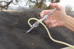
EBL can be transmitted through multi use blood-contaminated needles when vaccinating a herd.
Symptoms
Antibodies develop within two weeks of infection, but symptoms usually appear only after three years. Three symptomatic phases or condition types are observed:
- Cattle develop antibodies and symptoms are inconspicuous. The cattle remain infected for life and no clinical symptoms develop.
- Between 30% to 70% of infected animals develop permanent lymphocytosis (excess of a type of lymphocyte or white blood cell). This does not cause typical disease symptoms. Some cows remain in this preclinical stage for life with neither visible symptoms nor significant influence on performance. Affected cows may be culled due to poor performance caused by suppressed immunity. Animals with suppressed immunity experience more secondary infections, a decline in productivity and reproduction, and a shorter life expectancy.
- Very few (between 0,1% and 10%) of BLV infected animals develop tumours (lymphosarcoma) of the lymph glands and internal organs at between 4 years and 8 years. From 5% to 10% of clinically infected animals are peracute and die suddenly.
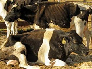
Animals with tumours usually have poor appetites (anorexia), emaciation and decreased milk production. Enlargement of one or more lymph nodes is the first visible sign. There may be visible lumps under the skin or in lymph nodes under the skin at the shoulder or above the udder. Widespread damage to lymph nodes and lymphoid tissue can occur and tumours can develop in the bone marrow, spleen, lungs, liver and uterus.
More than 50% of infected animals have anaemia. Cancer cells penetrate the bone marrow and suppress red blood cell formation. In the terminal stage, most animals develop leukaemia (excessive abnormal white blood cells). Other clinical symptoms include hindquarter paralysis, heart failure (sudden death), difficult breathing, blood in the dung and protruding eyes (exophthalmus). Such animals usually die within weeks or months of starvation, secondary bacterial infection, anaemia and vital organ failure.
Diagnosis
Vets can diagnose cattle on EBL clinical symptoms or a rectal examination (palpation) as the lymph nodes in the urogenital system are enlarged. Blood tests confirm the clinical diagnosis.Accurate tests can detect very small quantities of the antibody. Calves younger than 6 months can give a false positive test result as maternal antibodies may be present.
Prevention and Control
To date, there is no evidence that EBL is a zoonosis (a disease transmittable from animals to humans) or is detrimental to human health. However, EBL should be prevented and controlled due to the economic losses and under-performance associated with it. EBL-free countries ban animal imports from countries where it occurs.
Several European countries, including Denmark and Sweden, have eradicated EBL by testing and destroying infected animals. A positive EBL test must be followed by a positive test three to six months later before an animal is destroyed. If a herd tests negative every six months for two years, it can be declared EBL-free.
Note the following control measures:
- Infected herds should be isolated and quarantined with the ‘test and result’ method of control.
- Calves should not be fed blood-contaminated milk.
- Keep and rear calves separately, and handle the youngest first.
- As yet, there is no vaccine or effective treatment available for EBL infected animals.
- Animals bought in from auctions or other farmers should be EBL tested.
- Bio-security measures related to EBL should be strictly followed. Cattle test positive for antibodies two weeks (or more) after initial infection with the virus.
- When buying EBL-free cattle from a herd known to have EBL, quarantine the cows and re-test them after 21 days. If they test negative again, it is safe to transport them to a new destination farm. Consult a vet on the presence, impact and effect of EBL on a dairy herd when buying cattle.
As EBL is not a notifiable or controlled disease in South Africa, no official statistics are available on its prevalence and impact on the South African livestock industry, specifically the dairy industry. EBL may have entered SA by the import of infected cattle. Dairy cows in SA are mostly pasture-based with greater longevity than cows on total mixed rations (TMR).
The clinical effects of EBL occur only after three years and in pasture-based cows the longevity benefit is probably affected more than production or performance. Sporadic bovine leukosis (calf lymphoma or skin lymphoma) differs from EBL. Sporadic bovine leukosis occurs mainly in animals under 2 years old and is characterised by tumours in the skin and thymus. Only individual animals are affected. Sporadic bovine leukosis is thought not to be contagious, but limited information is available on this disease.
Contact Dr Jan du Preez on 012 843 5600 or email [email protected]


