

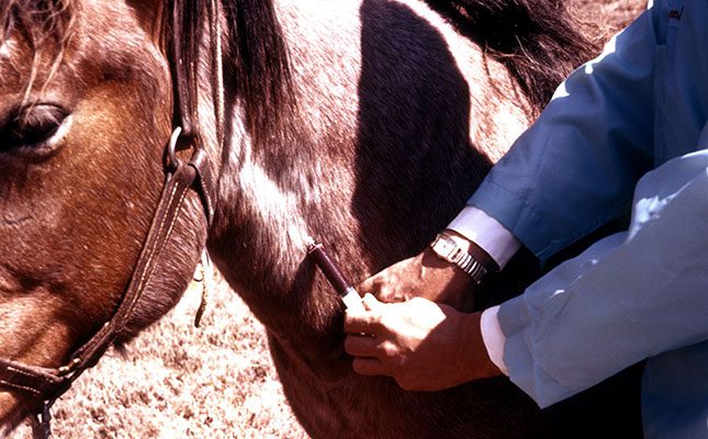
Aveterinarian called out to examine a horse will often insert a needle into the animal’s jugular vein and take blood samples. In most cases, two samples will be taken – in a red-topped and purple-topped tube respectively.
The former will be used for checking blood chemistry and diagnosing infectious diseases using serology (examination of blood serum), while the latter will be used to check the haematocrit and carry out blood smears. What does all this mean?
Different tubes, different tests
The red-topped tube contains no additives. The purple-topped tube contains an anticoagulant, which is a chemical that stops the blood from clotting.
If you take a sample from this unclotted blood for centrifugation, or even leave the tube standing upright, the red blood cells will settle at the bottom of the tube. Above them will be the serum, a golden-yellow liquid without blood cells or clotting factors.
The percentage of red blood cells to total blood volume is called the haematocrit or packed cell volume. It indicates whether the horse is anaemic (low haematocrit) or has an excessively high proportion of red blood cells (haemoconcentration).
The main cause of anaemia in horses is the presence of a blood parasite such as Babesia caballi or Theileria equi, which can cause babesiosis (biliary fever).
Haemoconcentration can be caused by any of a number of life-threatening conditions that need immediate veterinary intervention.
White blood cells
Another type of test is the blood smear, which assesses the proportion, number and shape of circulating white blood cells (leukocytes).
There are five types of leucocyte in the blood of horses (and all mammals), namely neutrophils, eosinophils, monocytes, lymphocytes and basophils. A high white cell count indicates a severe infection or allergy.
Platelets, which are produced in the bone marrow, are cells that are involved in blood clotting. Certain cancers, toxins and immune-related diseases can result in a deficiency of circulating platelets, a condition known as thrombocytopaenia.
Biochemical tests, which are used to identify bacteria and viruses, can also be conducted on blood.
When the blood sample is submitted for analysis, the accompanying document will list all the tests required. Each test will include the normal values and a diagnosis can be made by looking at how much the test values exceed or are lower than the norm.
For example, an increase in urea and creatinine levels will indicate a kidney problem, while an increase in bilirubin and alkaline phosphatase is associated with liver failure.
In the case of muscle disease or injury, the levels of creatinine kinase (CK) and aspartate aminotransferase (AST) become elevated within six to eight hours. CK occurs in high levels in skeletal and cardiac muscle.
In a horse, increased levels almost always signify acute muscle damage. Levels peak six to 12 hours after injury and can return to normal in three to four days. CK is often tested alongside AST when assessing muscle damage.
An increased level of blood lactate usually indicates that the horse has colic.
Blood can also be submitted to a laboratory for rapid identification (using polymerase chain reactions) of genetic patterns typical of hereditary diseases, as well as for pathogens such as African horse sickness.
Dr Mac is an academic, a practising equine veterinarian and a stud owner.


