

A tendon is a band of fibrous tissue that binds muscle to bone. Several bands of fibrous tissue are enclosed in a synovial tendon sheath and the contraction and extension of the tendon within this sheath is facilitated by synovial (lubricating) fluid.
There are two types of tendons in a horse’s leg: those that pull the legs forward and straighten out the joints (extensor tendons), and those that pull the legs backward and bend them (flexor tendons).
Most cases of tenosynovitis occur in the tendon sheaths surrounding the deep and superficial flexor tendons in three locations. Of these, the most commonly affected is the digital tendon sheath, which extends from just above the fetlock joint to the pastern joint, at the back of the legs.
The other two are the carpal sheath, which surrounds the flexor tendons at the back of the knee (carpus) on the forelimb, and the tarsal tendon sheath, which wraps around the flexor tendons at the back of the hock on each hind limb.
Tenosynovitis in the fetlock region can be due to rupture of either the deep or superficial flexor tendon, with secondary inflammation or chronic scarring extending into the tendon sheath. Inflammation can also occur if only the tendon sheath is torn or damaged.
Penetrating injuries
Swelling of the tendons can result in constriction by the annular ligament. This leads to interruption of the free flow of synovial fluid within the tendon sheaths and swelling above and below the ligament.
The tendon sheath lies just below the back of the fetlock; penetrating injuries that extend through the skin and tendon sheath into the tendon itself are fairly common.
This is often exacerbated by mud and dirt introduced between tendon and sheath. Although the initial wound is small, bacteria can proliferate within the sheath, causing swelling and, eventually, adhesions and scarring that extend up the tendons.
Treatment
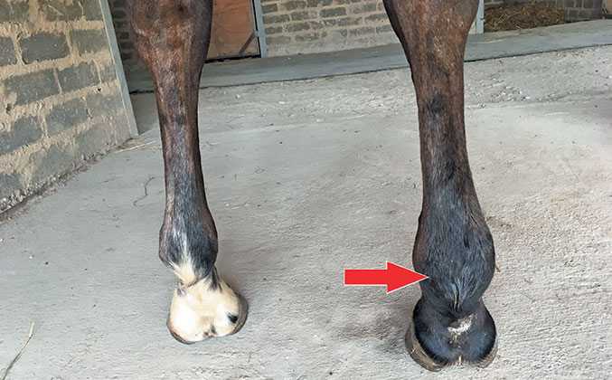
Early diagnosis and treatment are essential. The fetlock is obviously swollen and palpation of the joint and tendon sheaths is painful for the horse.
In more severe cases, adhesions may also be felt in the tendon sheath. Ultrasound and examination of synovial fluid can be used by your vet to aid in diagnosis.
Although the horse will not be particularly lame in the beginning, severe lameness will present within days if the horse is not treated. Cuts at the rear of the pastern have a poor prognosis, as infections in the synovial fluid are difficult to treat.
The entire leg should be soaked in Epsom salts and antibiotics injected for several days. Treatment with anti-inflammatories is also essential. The vet will sometimes inject cortisone into the sheath to reduce inflammation.
A farrier can apply wedged pads to reduce tension on the tendon, or your vet may surgically incise the annular ligament. Extracorporeal high-energy shock wave therapy has also been used successfully for treating tenosynovitis.
Dr Mac is an academic, a practising equine veterinarian and a stud owner.


