

Detecting fungus-infected maize early can help prevent contaminated grain from entering the food chain, reducing the risk of throat cancer linked to the consumption of infected maize products.In a recent study, a specialised camera was used to produce chemical images of maize kernels infected with fungi toxins. These toxins pose a threat to human and animal health. Through a process called chemical imaging, the camera takes photographs of contaminated kernels in infrared light. This light detects chemical changes caused by fungi long before any symptoms of infection are visible to the naked eye.
Each chemical and contaminant appears in a different colour, allowing it to be identified unmistakably. Chemical imaging thus makes it possible to distinguish between infected and healthy maize kernels in one rapid measurement. The researcher can then monitor the development of fungal growth in infected kernels over time. Whereas conventional microbiological techniques – which are complicated, labour-intensive and expensive – can take up to two weeks before positive identification is possible, chemical imaging allows individual kernels to be accurately sorted in seconds. Infected kernels can then be screened and removed before they enter the food chain.
It is crucial to find such grains as early as possible because maize, wheat, barley, oats and rice form part of the daily diet of millions of people. These products are consumed in raw or processed form for breakfast (porridges), lunch (bread), supper (pasta, rice and pap), or as an occasional snack. It is estimated that rice, maize and wheat provide at least 30% of the food calories for more than 4,5 billion people in 100 developing countries. Fungal infection of maize is therefore of global concern.
Maize is prone to infection by several fungi, especially Fusarium species. Fungal spores are everywhere and, in most cases, harmless. However, some spores can germinate, infiltrate grain, and grow under conditions such as rainfall shortly before harvest, or at high moisture and temperature levels during storage. The spores infect the leaves, stems, roots and cobs of the plant.
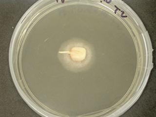
Growth rings
In the study, chemical imaging was able to distinguish three closely related, harmful Fusarium species. All three are capable of producing mycotoxins (fumonisins) that can cause throat cancer in humans through the consumption of infected grain products. These toxins also cause diseases in horses, pigs and rats.The chemical imaging allows researchers to visualise the radial growth rings of the three Fusarium species.
Similar to age rings in trees, these zones differ chemically, and it is possible, over time, to construct curves resembling microbial growth profiles. These curves can be painstakingly constructed using conventional microbiological techniques, but with chemical imaging the process can be achieved far more quickly using pixel counting and extrapolation.
Another advantage of chemical imaging is that it is safe and non-destructive, so has potential in silos or food processing factories. It can be used to test a variety of food products for similar fungi or spoilage bacteria. Chemical imaging can also be combined with conventional methods in food microbiology for rapid detection of foodborne pathogens.
Dr Williams is a postdoctoral research fellow at the Southern African Grain Laboratory in Pretoria. This article is based on his recent doctoral study in Food Science at Stellenbosch University.
Contact Dr Williams on 012 807 4019 or email [email protected].


