Enzootic bovine leukosis (EBL) is the most economically important oncogenic retroviral (cancer-causing) disease in dairy farming – and it starts with the bovine leukaemia virus (BLV).
BLV infection in herds varies between 1% to 100%. Infections are usually subclinical (asymptomatic). Mortality in severely infected dairy herds can be 5% or more per year.
All infected cattle develop antibodies and test positive for the virus throughout their lives, but only 30% develop persistent lymphocytosis (excess of lymphocytes; a lymphocyte is one of the white blood cells in the blood). Of these, 0,1% to 10% develop tumours.
Enzootic bovine leukosis occurs in several SA dairy herds. It’s neither a notifiable nor controlled animal disease under the Animal Disease Act of 1984. It is, however, a World Organisation for Animal Health-listed and notifiable disease. As SA is not EBL-free, cattle for export must test negative for the disease.
Three different manifestations of EBL are recognised: cattle with antibodies, but no clinical symptoms; cattle with persistent increased white blood cells (leucocytes); and cattle with solid lymphoid tumours.
Surveys in the US estimate that more than 44% of dairy and 10% of beef cattle are infected with the bovine leukaemia virus. Prevalence increases in larger herds. Sheep can also be infected with BLV.
In general, prevalence of viral infection increases with age. Enzootic bovine leukosis is characterised by a long incubation period of about three years (from infection until clinical symptoms are visible) that eventually results in the development of lymphosarcomas.
EBL should be distinguished from sporadic bovine leukosis (SBL). This occurs mainly in young cattle aged between four months and two years and mainly affects the skin and thymus gland.
SBL is not associated with EBL and is not contagious. Both occur in Southern Africa.
Infection
Cattle are infected with BLV at any age, including the embryonic stage, through the transfer of infected blood and blood products. Recently it was found that bovine udder epithelial cells may be infected with BLV as well.
In addition, BLV was found in the secretory epithelium of the human breast. This indicates a risk for the acquisition and proliferation of the virus in humans.
In fact, research (Prof Buehring et al, 2015) has established for the first time that BLV was significantly associated with human breast cancer.
The study does not identify how the virus infected the breast tissue. It could have been through consumption of unpasteurised milk or under-cooked meat.
As noted, bovine infection is caused by direct transmission of blood cells (lymphocytes) between infected and healthy animals (horizontal transmission). Small amounts of blood (100th of a blood drop, which cannot be observed with the naked eye) are enough to transmit infection.
The main modes of transmission include blood-contaminated needles, plastic gloves during pregnancy tests, ear tag applicators, tattooing/dehorning equipment and surgical instruments (iatrogenic transmission).
Transmission is also possible via blood transfusion and tests for tuberculosis, and mechanically by the bloodsucking insects Tabanus fuscicostatus (horseflies).
Clincial signs
There is currently no evidence that ticks can transmit the virus. Neither is there evidence that body fluids and excretions (saliva, faeces, urine, respiratory secretions, semen, uterine fluids) play any role in transmission and are considered to be non-infectious.
Congenital infection (calf infected in utero; vertical transmission) occurs in 4% to 8% of infected cows. Antibodies develop within two to five weeks after infection with BLV.
Symptoms in BLV-infected animals that are visible after three years include:
- Aleukaemic and clinically inapparent (unobtrusive) stage. This is the first stage. The majority of animals remain persistently infected with no outward signs of infection. Cattle develop antibodies and the symptoms are inconspicuous. They remain carriers for life. Results from some studies investigating the relationship between BLV and health, milk production and reproductive performance have been controversial.
- About 30% or more of the infected animals then develop permanent (persistent) lymphocytosis. This form of infection does not cause typical symptoms of the disease. Some cows remain in this preclinical disease stage for their entire production lifetime with no significant effect on their performance.However, many affected animals are culled due to poor milk production and reproduction performance caused by suppressed immunity. Animals with suppressed immunity have an increased incidence of secondary infections, variable increased somatic cell counts, a decrease in productivity and reproduction, and a shorter life expectancy. There is currently no convincing evidence that cattle with persistent lymphocytosis have an increased risk of developing lymphosarcoma.
- A small percentage (0,1% to 10%) of BLV-infected animals develops tumours (lymphosarcoma) when they are between four and eight years old. Approximately half of these animals have persistent lymphocytosis before developing lymphosarcoma.
Lymphosarcoma is rarely seen in animals younger than two years of age. In 5% to 10% of clinical cases the course is acute with affected animals dying suddenly without evidence of illness.
Clinical signs associated with the development of lymphosarcoma vary and are determined by the affected organ.
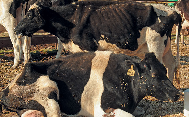
Lymphosarcoma can cause lesions in the spinal cords, heart, uterus, kidneys, spleen and liver. Animals with tumours usually have poor or no appetence (anorexia), emaciation (cachexia), weakness or general debility and decreased milk production.
Enlargement of one or more lymph nodes is the first visible sign.
More than 50% of sick animals have anaemia. Cancer cells penetrate the bone marrow and suppress the formation of red blood cells. In the terminal stage of the disease most animals develop leukaemia (excessive abnormal white blood cells).
Other clinical symptoms include paralysis of the hindquarter, heart failure (sudden death), difficult breathing, blood in faeces, abortion, spleen rupture, jaundice, abdominal pain, hydronephrosis and protruding eyes (exophthalmus).
Animals affected in this way usually die within weeks or months as a result of starvation, secondary bacterial infections, anaemia and failure of vital organs.
Increased culling rates and a greater susceptibility to other infectious diseases (such as mastitis, diarrhoea and pneumonia) have also been demonstrated among dairy cattle that test positive for BLV.
Therefore, despite no obvious clinical signs during the long subclinical infection period, economic losses caused by persistent BLV infections can be great.
Serious consequences
When compared to dairy cattle herds with no BLV infection, those with infected cows yielded 218kg (3%) less milk annually calculated across the entire dairy industry in the US. BLV resulted in a loss to producers of US$285 million (about R2 850 million) due to reduced milk production in BLV-positive herds.
In addition:
- The presence of BLV-positive cows in some dairy herds led to a slight, but statistically significant increase in calving intervals.
- The somatic cell (pus) count is higher in BLV-positive cows.
- In dairying, the somatic cell count is an indicator of the quality of milk – and the lower, the better.
- In some dairy herds culling rates increased with age and were significantly greater in seropositive than in seronegative cows.
- Survival analysis of cows 3,5 years and older showed BLV-negative cows lived longer.
- Genetic merits are connected to the susceptibility of dairy cows to BLV infection. Cows that are genetically high milk producers are more susceptible to BLV infection.
- To reiterate, most BLV-infected dairy cows exhibit a reduced economic value expressed in higher susceptibility to infectious diseases, decreased productivity, lower reproduction and higher culling rate.
- Because BLV infection affects the immune system of cattle, its impact on herd health and economy could be more extensive than direct loss from death following lymphomas (lymphosarcomas).
In addition, the detrimental effect of BLV infection on the bovine immune system may cause a higher susceptibility to opportunistic and infectious agents resulting in clinical diseases (such as mastitis, metritis, lameness diseases and ringworm (Trichophyton verrucosum) infection).
Also, as noted, the most pronounced clinical manifestation of BLV infection is the appearance of lymphomas with 100% mortality. A US study estimates that lymphoma cases cost the US dairy industry more than US$16 million (R230 million) annually.
This includes direct costs resulting from loss of diseased cows. The cost of vet services, medications and time span until the right diagnosis is determined, may make this figure much higher.
Moreover, these costs do not include those resulting from the ban on exportation of livestock, as well as semen and embryos from affected dairy herds to countries worldwide.
Diagnosis
Veterinarians can often diagnose cattle on the clinical symptoms of EBL. Enzootic bovine leukosis can also be diagnosed with a rectal examination (palpation), as the lymph nodes in the urogenital system are enlarged.
A clinical diagnosis must, however, be confirmed with histology, virology and serology, ELISA and PCR (blood tests). Leukosis virus antibodies in individual or pooled milk samples can also be used for diagnostic purposes.
Positive serology or virology for BLV confirms viral infection but not the presence of lymphosarcoma. The diagnosis of lymphosarcoma must be made by cytology and histopathology.
Prevention and control
Economic losses and under-performance are important reasons why EBL should be prevented and controlled. Some countries free from EBL do not allow importation from countries where the infection occurs. Again as noted, research has established for the first time that BLV is significantly associated with human breast cancer. Further research will determine if EBL is a zoonosis disease (transmittable from animals to humans).
In several European countries, EBL has been eradicated through the testing and culling of infected animals.
Positive EBL testing of herds must be followed up by further tests between three to six months before culling.
Only if a herd is tested every six months for a period of two years, and the test results remain negative, can a herd be declared free from EBL.
Infected herds should be kept in isolation and quarantine with the ‘test and culling’ method of control.
Calves should not be given milk contaminated with blood. They must be kept and reared separately. The youngest calves should always be handled first.
The most commonly recommended eradication protocol, ‘test and culling’) involves:
- Identifying infected animals using a serologic test;
- Culling seropositive animals immediately;
- Retesting the herd in 30 to 60 days;
Using PCR to test young calves and as a complementary test for clarifying test results in herds with a low prevalence of infection. Testing and culling is repeated until the entire herd tests negative.
There are no vaccines or effective treatment for animals affected with EBL. Animals should be tested for EBL before auctions or direct buying from cattle farmers, and all biosecurity measures related to EBL should be followed.
Cattle only test positive for antibodies two to five weeks after infection with EBL virus. Therefore, if negative EBL-tested cattle are purchased from a positive EBL-tested herd, the cattle should be kept in quarantine and tested again after 21 days.
If all cattle test negative for the second time it’s safe to take them to your farm.
Vets should be consulted about the prevalence, impact and effect of EBL on dairy herds when acquiring cattle.
Eliminating the movement of blood from infected animals to healthy animals is the cornerstone of prevention protocols.
In calves, feeding colostrum from seronegative cows is often advocated.
However, most epidemiologic evidence suggests that the protective effect of colostral antibodies outweighs the risk of infections, particularly in high-prevalence herds.
Replacing whole milk feeding with high-quality milk replacer may also be considered. Bloody milk should never be fed to calves.
Bloodless methods of dehorning should be employed. Equipment used for castration, tattooing, ear-tagging or implanting should be properly cleaned and disinfected between use on animals if they are expected to live past two years of age.
Transmission can be decreased in adult dairy cows or heifers by using a seperate new rectal sleeve for each pregnancy test.
Artificial insemination or embryo transfer (using negative recipients) may limit transmission.
In beef herds, the use of a negative bull may limit transmission too, but natural service is an uncommon method of viral transmission unless breeding is traumatic.
Recommendations
Recommendations regardless of animal age include disinfection of equipment that has come into contact with blood or body tissue. Single-use, disposable needles should always be used for blood collection and intramuscular injections.
Single-use disposable needles are also better for vaccinations, but the risk of transmitting BLV virus via subcutaneous vaccination is low.
Handling facilities that become contaminated with blood should be cleaned between use on animals. Fly control will help minimise the potential for tabanid-associated transmission. Blood transfusions and vaccines containing blood, such as those used for babesiosis and anaplasmosis, are particularly potent means of spreading the disease, and donors must be carefully screened.
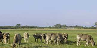
Most dairy cattle in SA are kept on pasture and their lifespan is longer than cattle on a total mixed ration (TMR). The clinical effects of the disease only occur after three years and it is believed that infected dairy cattle on pasture are likely to be more badly affected when it comes to production and performance (because of their longevity).
Phone Dr Jan du Preez on 083 656 3638 or email [email protected].

