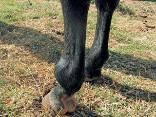
The fetlock joint is made up of the lower end of the cannon bone and the top of the pastern bone. Two small bones, the sesamoids, lie at the back of the joint. These cradle the flexor tendons and are attached to both the suspensory and sesamoid ligaments.
Both the front and hind fetlocks act as shock absorbers when a horse moves. When the legs become tired, the fetlocks are at high risk of being injured.
Cartilage covers the ends of the bones in every joint. As the joint flexes and extends, each cartilage surface moves against the other, lubricated by joint fluid and encased in a joint capsule. Over the years, the cartilage becomes damaged and worn away. As the body attempts to repair the damage, inflammation occurs and bone is built up around the damaged cartilage. This is called osteoarthritis.
In older horses particularly, osteoarthritis of the fetlock joint often occurs as a result of small injuries over time. It is more common in the front legs as they carry 60% of the horse’s weight.
During the acute phase of injury, the fetlock is often puffy and painful, and the horse is severely lame. Later, the entire joint becomes swollen and hardened. In many cases, the chronic damage is no longer painful and the horse may eventually become fairly sound and can be ridden again.
Diagnosis
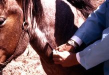
Vets commonly use a flexion test to check for osteoarthritis of the fetlock joint. Riders often try to carry out the test themselves when they notice the hardened swelling. But rough handling of a damaged fetlock can cause severe lameness, and flexion tests are best left to a professional. The vet may use radiography (a good idea) or take samples of the synovial fluid to assess the state of the cartilage more accurately.
Treatment
In the early stages of cartilage damage, the affected leg can be put in a bucket of ice to reduce pain and inflammation. Your vet may also prescribe injectable and oral pain killers or anti-inflammatories. In some cases, hyaluronic acid is injected directly into the joint.
Occasionally, X-rays show chips or small fractures in and around the fetlock joint and surgery is indicated. The chance that your horse will be able to continue its previous level of work depends on what type of damage is found on X-rays, MRI or during surgery.
Even if it recovers fully, you should use support bandages or padded boots to protect the fetlock as the horse is brought back into work.
Chronic cases
In old horses, where the bone has already grown around the joint and the horse is no longer lame, there is a good chance it will remain sound.
It is not necessary, and may even be uncomfortable for the horse, to use bandages and boots in these chronic cases.
During winter, the horse may be fairly stiff early in the morning, but will warm up and move more freely after it is exercised.
You can massage horse liniment around the swollen joint. Glucosamine, MSM and chondroitin powder should be given daily in food over the long term, as this combination supports cartilage regeneration.
Dr Mac is an academic, a practising equine veterinarian and a stud owner.


